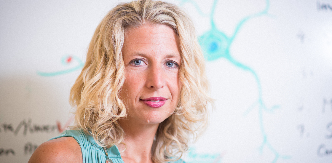
Beth Stevens: Casting immune cells as brain sculptors
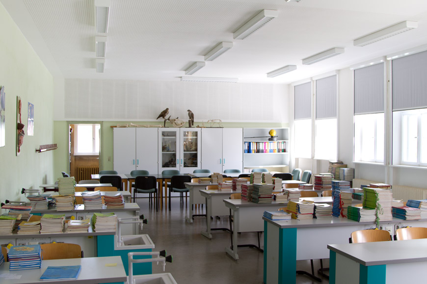
Shortly after Beth Stevens launched her lab at Boston Children’s Hospital in 2008, she invited students from the Newton Montessori School, in a nearby suburb, to come for a visit. The children peered at mouse and rat brains bobbing in fluid-filled jars. They also learned how to position delicate slices of brain tissue on glass slides and inspect them with a microscope.
This visit sparked a running relationship with the school, with a steady stream of students visiting the growing lab each year. Soon it became too complicated to bring so many children to the lab, so Stevens decided to take her neuroscience lessons on the road, visiting a number of local elementary schools each year. Last year, she dropped in on the classrooms of her 5- and 8-year-old daughters, Zoe and Riley.
“The kids got really excited,” Stevens says. “It’s become such a thing that the principal wants me to come back for the whole school.”
Stevens’ enthusiasm for science has left a lasting impression on researchers, too. Her pioneering work points to a surprise role in brain development for microglia, a type of cell once considered to simply be the brain’s immune defense system, cleaning up cellular debris, damaged tissue and pathogens. But thanks to Stevens, researchers now appreciate that these non-neuronal cells also play a critical role in shaping brain circuits.
In a 2012 discovery that created a buzz among autism researchers, Stevens and her colleagues discovered that microglia prune neuronal connections, called synapses, in the developing mouse brain. The trimming of synapses is thought to go awry in autism. And indeed, emerging work from Stevens’ lab hints at a role for microglia in the disorder.
Stevens has already earned praise and several prizes for her work. In 2012, she received the Presidential Early Career Award for Scientists and Engineers, the most prestigious award that the U.S. government bestows on young scientists. And in October, she’ll deliver one of four presidential lectures at the world’s largest gathering of neuroscientists — the annual meeting of the Society for Neuroscience — an honor she shares with three neuroscience heavyweights, including two Nobel laureates.
“The field is probably expecting a lot from Beth,” says Jonathan Kipnis, professor of neuroscience at the University of Virginia. Stevens has put microglia at the forefront, Kipnis says. “What used to be a stepchild of neuroscience research is now getting a lot of attention, and I think in part it’s due to her research.”
Curious mind:
Stevens was born in 1970 in Brockton, Massachusetts, where her mother taught elementary school and her father was the school’s principal. As a child, she was deeply inquisitive, eager to understand how things work. She enjoyed collecting bugs and worms, and would analyze these precious specimens in makeshift labs in her backyard.
But a career in science wasn’t on her radar until high school, when she took a biology class with an inspiring teacher named Anthony Cabral. “He totally made me realize that this could be a career, that I could be a scientist,” Stevens says. “It was that one class that changed it, and I’m like, ‘Okay, I’m going to do this.’”
In 1988, she began studying biology at Northeastern University in Boston, which offered an unusual opportunity. It had a unique cooperative education program that allowed Stevens to spend several semesters working full time in medical labs after finishing her coursework.
After that experience, Stevens knew she wanted to find a job in a research lab. After graduating in 1993, she joined her then-boyfriend Rob Graham, now her husband, in Washington, D.C., where he had landed a job in the U.S. Senate. Stevens headed to the National Institutes of Health (NIH) in Rockville, Maryland, to apply for a job as a research assistant.
At around the same time, neuroscientist R. Douglas Fields was launching his lab at the NIH. He studied how neural impulses influence glia — a class of non-neuronal cells that includes microglia — and shape the structure of the developing brain. Fields readily hired Stevens despite her lack of expertise in neuroscience. “I was impressed with her work ethic, energy and drive,” he says.
Stimulating research:
In Fields’ lab, Stevens used a multi-compartment cell culture system to investigate whether stimulating neurons influences the activity of Schwann cells, glial cells that produce a fatty substance called myelin, which insulates nerves1. She discovered that patterns of neural impulses similar to those that occur during early development influence the maturation of Schwann cells and the production of myelin.
The findings added to mounting evidence that glia and neurons communicate with each other, a newly emerging concept at the time.
“What I loved about the glia research was that there were so few neuroscientists studying it; it was such a mysterious part of neuroscience,” Stevens says. “Those years in Doug’s lab were really exciting because it was a new field.”
Stevens spent five years in Fields’ lab. “She was doing extraordinary work,” Fields says. “She had the potential and the interest to do neuroscience research, and I recommended that she should consider going to graduate school.”
But Stevens didn’t want to give up her position in the lab, and at that time, the NIH did not allow its researchers to have graduate students. So she and Fields convinced the University of Maryland, College Park, just 10 miles away, to allow her to take graduate courses in neuroscience while completing the necessary research for her Ph.D. in Fields’ lab.
In 2000, less than two years after starting graduate school, Stevens published a paper in Science showing that nerves in the peripheral nervous system (located outside the brain and spinal cord) use chemical signals to communicate with Schwann cells2. Two years later, she reported in Neuron that a similar form of communication occurs in the brain, between neurons and oligodendrocytes, the myelin-producing cells in the brain3.
As she was closing in on her Ph.D., Stevens sought career advice from Story Landis, then-director of the National Institute of Neurological Disorders and Stroke. Landis turned Stevens on to the possibility of starting her own lab one day. “I convinced her that she really had the abilities and energy and intelligence to run an independent research program,” Landis says.
In 2004, Stevens sought a postdoctoral fellowship with neurobiologist Ben Barres at Stanford University. “She was already seen as a leading researcher in the glial field,” recalls Barres, who promptly hired her. “She had done all sorts of beautiful work on glia.”
In Barres’ lab, Stevens continued to explore the dialogue between neurons and glia, turning her attention to star-shaped glia called astrocytes. Barres and his team had discovered that astrocytes help neurons form synapses4. To get a better handle on this process, Stevens examined how astrocytes influence gene expression in neurons in the developing mouse brain.
To her surprise, she found that astrocytes trigger neurons to produce a ‘complement’ protein that is best known for its role in the immune system. There, the protein serves as an ‘eat me’ signal, flagging pathogens and debris for removal. She found that neurons deposit this tag around immature synapses, but not mature ones, in mouse brain tissue, and mice that lack this protein have too many immature synapses. The findings suggested that astrocytes might help eliminate synapses by triggering the complement cascade5.
But it was still unclear exactly how the tagged synapses are cleared. The prime suspects were microglia, the only cells in the brain known to have the receptor for the ‘eat me’ signal.
Stevens set out to test this hypothesis in her own lab: After four years as a postdoc, she had decided to branch out on her own. In 2008, neuroscientist Michael Greenberg — chair of the neurobiology department at Harvard — recruited her to the Harvard-affiliated Boston Children’s Hospital. Even when her lab was in its infancy, she had little trouble convincing new staff to join her.
“A lot of people might be a little hesitant to join a new lab,” says Dorothy Schafer, a former postdoctoral fellow in Stevens’ lab who is now assistant professor of neurobiology at the University of Massachusetts-Worcester. “But I was so excited by the research, and she was so energetic and extremely positive, and just seemed like a very nice person.”
One decision Stevens made early on was to continue to studying microglia in mice rather than experiment with new model systems. “You’ll never see her working on songbirds, because she has this aversion to birds,” Schafer says. “I think they think her curly blond hair is a nest or something, and she’s had really bad experiences with many types of birds dive-bombing her head.”
Microglia maven:
Just four years into her foray as an independent researcher, Stevens found the proof she had been looking for. In 2012, her team published evidence that microglia eat synapses, especially those that are weak and unused6.
The findings pinned down a new role for microglia in wiring the brain. They also helped to explain how the brain, which starts out with a surplus of neurons, trims some of the excess away. Neuron named the paper its most influential publication of 2012.
Stevens continues to study the function of microglia in the healthy brain, most recently uncovering preliminary evidence that a certain protein serves as a ‘don’t eat me’ tag that protects synapses from being engulfed by microglia. She is also exploring the role of microglia in disorders such as autism.
Several studies suggest that microglia are more active and more numerous in the brains of people with autism than in controls. Stevens and her team are looking at whether the activity of microglia is altered during brain development in mouse models of autism.
Research on microglia has exploded in the past decade, Stevens says, but there is still much to be done. “It’s so wide open,” she says. “Nothing is known about [microglia] except for a few things. That’s a whole career’s worth of stuff to do.”
And there should be plenty left over for any ambitious children she inspires, too.
This article was originally published on Beth Stevens: Casting immune cells as brain sculptors


